The US technique - 2
The primary ultrasound (US) landmark is the ASIS (Fig 1).
This has a triangular shape with its apex superficial and, on US imaging, has a hyperechoic (white) demarcation with an anechoic (black) filling. Lying on the medial surface of the iliac bone is the iliacus muscle.
By gently moving the probe caudad and cephalad the three lateral abdominal muscles can be identified.
Deep to the muscles is the peritoneum and viscera. Peristalsis is usually observed.
The nerves are usually found between the innermost muscle layers and are seen as hypoechoic ellipses.
Click here to play a video of the US-guided ilioinguinal nerve block.
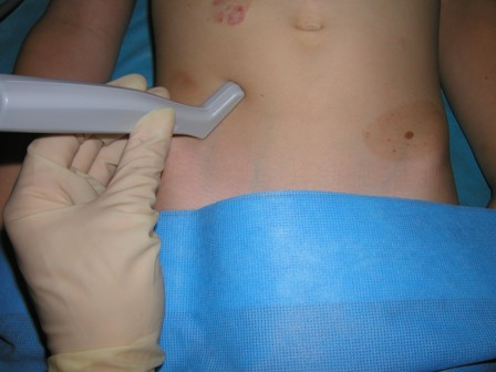
Fig 1 Hockey stick probe, the heel of which is resting on the ASIS. An in-plane needling technique is shown
Fig 2 The US-guided ilioinguinal nerve block technique