Treating Life-threatening Injuries
To identify and treat life-threatening injuries:
Signs of tension pneumothorax:
- Respiratory distress, hypoxia, tachycardia and hypotension
- Reduced chest movement, reduced breath sounds, resonant to percussion on the affected side
- Tracheal deviation away from the affected side is a late sign
- Neck veins may be distended, unless hypovolaemic
Treatment of pneumothorax is open thoracostomy:
This procedure can be performed rapidly. This must be followed by a chest drain via the thoracostomy in spontaneously breathing patients. There is no need to insert a chest drain immediately in ventilated patients.
Needle decompression can be used in spontaneously breathing patients with haemodynamic instability or severe respiratory compromise if the expertise or equipment necessary to insert a chest drain is not immediately available:
- If available use a purpose-made thoracocentesis device with a long needle. Otherwise a 14G cannula can be used, but this will not reach the pleural space in 50% of cases and kinks easily
- Insert the cannula attached to a syringe in the 2nd intercostal space in the mid-clavicular line, just above the rib
- Advance slowly until air is aspirated
- Remove the syringe and needle, leaving the cannula in place. There may be a hiss of air
- This is a temporary measure and does not completely re-expand the lung. Insert a chest drain as soon as possible
Open pneumothorax
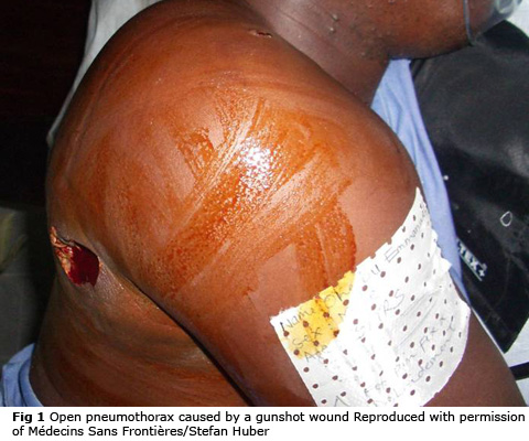
An open pneumothorax (Fig 1) or sucking chest wound allows air to pass through the wound instead of down the trachea into the lungs.
Treatment of open pneumothorax is to place an occlusive dressing over the wound, taped on three sides to act as a one-way valve allowing air out of the pleural space but not in. Purpose-made chest seals are available.
A chest drain will be required later.
Massive haemothorax
Massive haemothorax is defined as >1500 ml blood in the chest.
Signs of massive haemothorax:
- Reduced chest movement
- Reduced breath sounds
- Dullness to percussion on the affected side
- Signs of shock
Treatment of massive haemothorax:
- Gain IV access, have warmed blood available
- Insert a chest drain
Usually, bleeding stops following chest drain insertion as the lung re-expands. Rarely, a haemothorax of greater than 1500-2000 ml or continued bleeding of more than 200-300 ml/hour may be an indication for thoracotomy.
This is a high-risk procedure in a resource-poor setting and requires post-op critical care.
Flail chest
Flail chest occurs when multiple rib fractures allow a segment of the chest wall to move the wrong way. This impairs ventilation.
Underlying lung contusions will also lead to hypoxia.
Poorly managed pain leads to retention of secretions and pneumonia.
Initial management of flail chest is oxygen, good analgesia and positioning
Cautiously titrated IV morphine or low dose ketamine may be required. Don't forget regular paracetamol.
Regional analgesia using a thoracic epidural or paravertebral blocks are very effective if expertise are available. Intercostal blocks, intrapleural local anaesthetic and lignocaine patches are less effective, may be easier to use but also have potential complications.
Patients with chest injuries are likely to be more comfortable and breathe better sitting up. Clear the spine as early as possible. If the spine cannot be cleared tilt the bed head-up. A careful balance of risks is required - the risk of ventilatory failure may outweigh the risk of exacerbating a potential spinal injury.
Patients need close observation, humidified oxygen if possible and chest physio (with good analgesia!).
Intubation and ventilation may be required. Non-invasive ventilation may be of benefit in selected patients.
Pneumothorax is likely, even if it is not obvious initially.
A chest drain is likely to be necessary, particularly in patients needing positive pressure ventilation.
Cardiac tamponade
Cardiac tamponade causes circulatory compromise, but may be identified when examining the chest.
It should be suspected in penetrating injuries to the chest and abdomen and can also occur in blunt trauma.
Signs:
- Hypotension
- Tachycardia
- Distended neck veins
- Muffled heart sounds
Treatment of cardiac tamponade requires emergency thoracotomy.
In witnessed cardiac arrest from penetrating chest trauma, resuscitative thoracotomy in the emergency department may be indicated.
Pericardiocentesis may buy some time if thoracotomy is not immediately available.