Anatomical Variation
Ultrasound imaging used to identify the internal jugular vein during cannulation has shown how variable its anatomical position can be relative to the carotid artery.
The vein is normally lateral to the artery, but in some patients may overlie the artery, or even, though rarely, be medial.
Fig 1 shows an ultrasound scan of the right internal jugular vein (IJV) and common carotid artery (CCA) in the neck.
Fig 2 shows the possible relationships between these structures, with their frequency within the population.
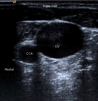
Fig 1 Ultrasound scan of the internal jugular vein and common carotid artery in the neck
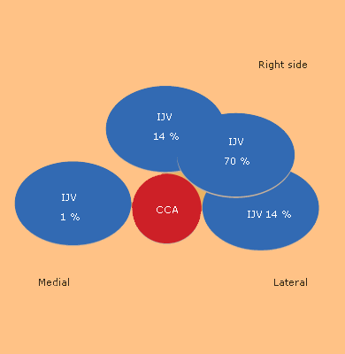
Fig 2 The possible relationships between the CCA and IJV (% of individuals)